


The Needle's Eye View
Science might be catching up to sci-fi, thanks to 'smart needle' technology developed by OU biomedical researchers.
Surgeons have been using cameras to look inside their patients’ bodies for generations, but the procedure has never been easy and potential risks can be substantial. Now, two University of Oklahoma biomedical researchers are on the cusp of a breakthrough technology that could make endoscopy safer and more useful than ever.
OU professors Qinggong Tang and Chongle Pan have been collaborating for several years on a big project, but for them to be successful, they’ve had to think small—so small that they’re pushing the boundaries of science and technology to get there. They say there is much work left to do, but their successes have already drawn the attention of the National Science Foundation and the Oklahoma Center for the Advancement of Science and Technology, which recently awarded Tang a $500,000 NSF CAREER Award and a $135,000 OCAST Oklahoma Health Research Award, respectively.
“This funding will help us advance an endoscopic platform,” Tang says. “Our long-term goal is to put the platform into clinics. We aren’t quite there yet, but such support will allow us to push through technology and lab testing before we reach clinical trials.”
Tang’s vision for the novel, optical technology utilizing a minute imaging device includes broad applications within the human body. His initial focus is on the tiny, but problematic, epidural spaces in human spinal cords where doctors use needles to administer anesthesia, steroids and other pain medicine. Hazards include piercing blood vessels and nerves, and there is always the danger of missing the target area entirely, injecting drugs into blood vessels or other tissue, and triggering a host of complications.
Today, doctors go by experience and their sense of feel to get the tips of needles where they need to go, but precision can be a problem. Depending on where on the spine a doctor is working, accuracy can range from 50 to 80%, Tang says.
“Over 20% of the time, they miss,” he says. “Either they put the drug in the wrong place or the needle damages unintended tissues, causing chronic headaches that can last for months.”
If doctors had an instrument that would allow them to see where their needles are going, they could avoid trouble before causing damage. That was the challenge Tang and Pan embraced when they began their research five years ago.
Tang, an assistant professor of biomedical engineering, and Pan, an associate professor of microbiology and computer science, are harnessing the power of artificial intelligence, or AI, to refine what could soon make common surgical procedures safer and more effective.
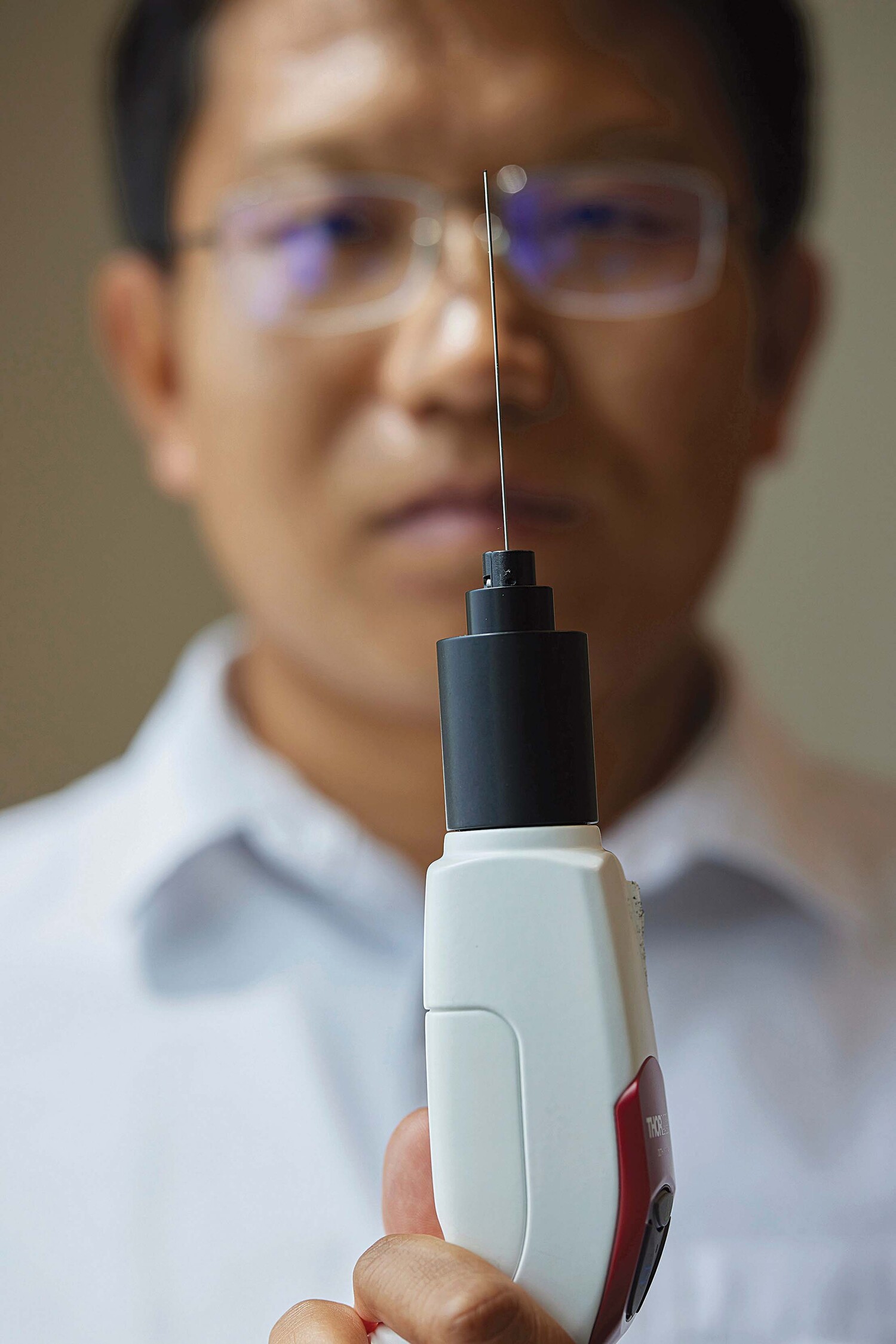
They have designed the “smart needle,” a camera smaller than the depth of a credit card that fits inside the hollow bore of an epidural needle. The device will allow doctors to find reliable pathways through the body by acquiring tissue images and data never seen before. While the platform is initially being designed for reaching the epidural space in spinal cords, Tang says the technology will do much more than that.
Eventually, doctors could use such technology to guide surgical tools, he says. In addition, the system could break up kidney stones and offer a better way to collect biopsy tissue from kidneys, livers, prostate glands and other organs.

As a computer scientist, Pan is employing AI to teach the system how to identify tissue within the body before the endoscopic needle reaches it. Through AI, doctors will be alerted when their needles approach nerves, blood vessels and other vulnerable tissues, he says.
“It is a long and arduous process to develop a ‘deep learning’ model by training it with tens of thousands of images from blood vessels, muscle, cartilage, fat and other tissue types to ensure accurate recognition,” he says.
“It’s a little like driver-assist for the surgeon,” Pan adds. “Instead of guiding a car down a highway, this system may soon help guide an endoscopic needle along a safer path through the human body.”
Chip Minty is a Norman-based writer and the principal of Minty Communications LLC.
To comment on this story, click here.